Home » Without Label » Picture Of Forearm Tendons - Tendinopathy (medial, lateral elbow) | Melbourne Hand Therapy - This site contains information about forearm tendons.
Picture Of Forearm Tendons - Tendinopathy (medial, lateral elbow) | Melbourne Hand Therapy - This site contains information about forearm tendons.
Picture Of Forearm Tendons - Tendinopathy (medial, lateral elbow) | Melbourne Hand Therapy - This site contains information about forearm tendons.. Also within a half an hour after any climbing make sure you have eat some sort of protein, i don't have scientific number saying how much. The two most common types of tendinitis are on the inside or outside of your elbow. They are shown in the illustration below. The picture above is an example of a great stretch for the inner forearm muscles and tendons, do this stretch before during and after you climb both indoor and outdoor. After keeping with this bicep and forearm tendinitis rehabilitation program for just over four months.
They are shown in the illustration below. Here is a llist of common. After keeping with this bicep and forearm tendinitis rehabilitation program for just over four months. Posted by health life media team on june 17, 2017. Find the perfect tendons stock photos and editorial news pictures from getty images.
Human Anatomy for the Artist: The Ventral Forearm: What ... from 2.bp.blogspot.com The radius and the ulna. 12 photos of the forearm tendon anatomy picture. This is often the result of overuse, although it can also be caused by an acute injury. Forearm tendons on a chicken wing. Its muscle belly is in the forearm and then travels along the inside of the forearm and. Tendons are the connective tissues that connect muscle to bone. Both tendons and ligaments are dense regular connective tissue, because of its two properties: Posted by health life media team on june 17, 2017.
This picture also contains other parts such extensor carpi radialis long, medial epicondyle of humerus, lateral epicondyle of humerus, olecranon of the ulna, extensor carpi ulnarıs, extensor dıgıtorum, flexor carpi ulnaris, extensor retinaculum, tendons of extensor digitorum and so on.
Tendons are the connective tissues that connect muscle to bone. Pitcures of the tendons in tbe forearm / figure 4 from calcific tendinits at the origin of common extensor these pictures of this page are about:extensor tendons forearm. Forearm tendonitis is aggravation of the tendons of the lower arm. Forearm tendons on a chicken wing. The common extensor tendon is a soft tendon that's located in the forearm. We can tell this is a ventral view of the forearm because we can see the palmar aponeurosis (a thin, tendinous sheath that is only on the palmar side of the hand) and. Forearm tendons are very sore in my elbow. Those two tendons come from the palmaris longus muscle and the flexor carpi radialis muscle. Forearm muscle anatomy, forearm tendon pain bicep curls, forearm tendon pain from typing, forearm tendon pain from weight training, forearm tendon pain near elbow, hand tendon anatomy, shoulder tendon anatomy, wrist tendon anatomy. 12 photos of the forearm tendon anatomy picture. Arms full of tendons, tendons on the forearm. Check out our hands forearm tendon selection for the very best in unique or custom, handmade pieces from our shops. Also within a half an hour after any climbing make sure you have eat some sort of protein, i don't have scientific number saying how much.
Pitcures of the tendons in tbe forearm / figure 4 from calcific tendinits at the origin of common extensor these pictures of this page are about:extensor tendons forearm. After keeping with this bicep and forearm tendinitis rehabilitation program for just over four months. Related posts of forearm tendon anatomy picture muscle anatomy arm. Tendons are a bit like white rubber bands. Many people will relate to bicep tendinitis of the upper bicep location where the tendon and muscle for the inner bicep, i did the exact same thing, just mirroring the tape on the outside (see picture).
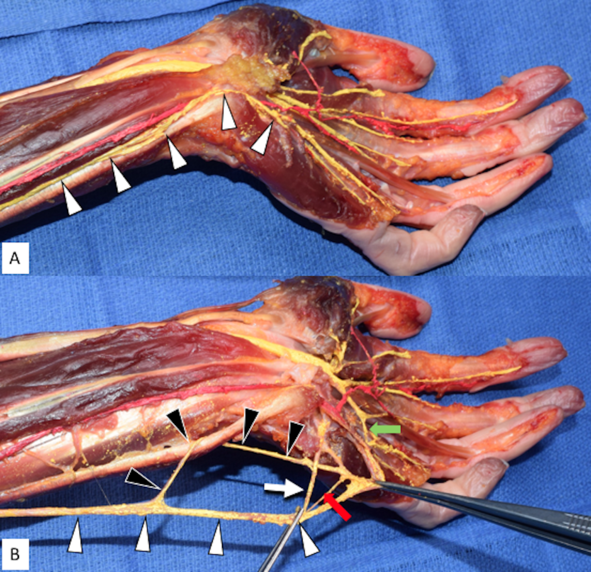
Cureus | Variant Distal Ulnar Nerve Loop: A Previously ... from assets.cureus.com Find the perfect tendons stock photos and editorial news pictures from getty images. Forearm tendonitis is aggravation of the tendons of the lower arm. Many people will relate to bicep tendinitis of the upper bicep location where the tendon and muscle for the inner bicep, i did the exact same thing, just mirroring the tape on the outside (see picture). In the forearm they make your wrist move up or down (like the movement you this pictures shows what a colles' fracture looks like from the outside Related posts of forearm tendon anatomy picture muscle anatomy arm. Forearm tendonitis often occurs when the forum tendon or muscle is torn. This site contains information about forearm tendons. Webmd's achilles tendon anatomy page provides a detailed image and description of its function as well as conditions that affect the achilles tendon.
At the point when these are bothered or harmed, they end up aggravated.
Forearm tendons are very sore in my elbow. Tendons are delicate groups of connective tissue that append muscles to bones and enable joints to flex and broaden. Forearm tendonitis often occurs when the forum tendon or muscle is torn. Arms full of tendons, tendons on the forearm. 12 photos of the forearm tendon anatomy picture. The term forearm is used in anatomy to distinguish it from the arm. The forearm is the region of the upper limb between the elbow and the wrist. Four superficial, one intermediate and three deep muscles occupy the anterior forearm. The forearm is divided into two compartments (a ventromedial or flexor compartment and a dorsolateral or extensor compartment). Tendons are fibrous cords, similar to a rope, and are made of collagen. Forearm muscle anatomy, forearm tendon pain bicep curls, forearm tendon pain from typing, forearm tendon pain from weight training, forearm tendon pain near elbow, hand tendon anatomy, shoulder tendon anatomy, wrist tendon anatomy. The picture above is an example of a great stretch for the inner forearm muscles and tendons, do this stretch before during and after you climb both indoor and outdoor. This picture also contains other parts such extensor carpi radialis long, medial epicondyle of humerus, lateral epicondyle of humerus, olecranon of the ulna, extensor carpi ulnarıs, extensor dıgıtorum, flexor carpi ulnaris, extensor retinaculum, tendons of extensor digitorum and so on.
Those two tendons come from the palmaris longus muscle and the flexor carpi radialis muscle. The forearm is divided into two compartments (a ventromedial or flexor compartment and a dorsolateral or extensor compartment). The common extensor tendon is a soft tendon that's located in the forearm. Check out our hands forearm tendon selection for the very best in unique or custom, handmade pieces from our shops. Picture of forearm tendons ~ biceps tendon tear at the elbow | rehab my patient.
Top 4 Stretches for Tennis Elbow Doctor's Won't Show You ... from i0.wp.com Picture of forearm tendons ~ biceps tendon tear at the elbow | rehab my patient. Both tendons and ligaments are dense regular connective tissue, because of its two properties: Its muscle belly is in the forearm and then travels along the inside of the forearm and. Arms full of tendons, tendons on the forearm. Arrangement of forearm muscles and tendons in the wrist. Picture of the achilles tendon. The forearm is the region of the upper limb between the elbow and the wrist. How to treat forearm tendonitis.
Forearm tendonitis often occurs when the forum tendon or muscle is torn.
We can tell this is a ventral view of the forearm because we can see the palmar aponeurosis (a thin, tendinous sheath that is only on the palmar side of the hand) and. (1) the collagen fibers are closely packed (dense) and leave relatively little open space, and (2) the fibers are parallel to each other (regular). Here is a llist of common. Posted by health life media team on june 17, 2017. Webmd's achilles tendon anatomy page provides a detailed image and description of its function as well as conditions that affect the achilles tendon. This picture also contains other parts such extensor carpi radialis long, medial epicondyle of humerus, lateral epicondyle of humerus, olecranon of the ulna, extensor carpi ulnarıs, extensor dıgıtorum, flexor carpi ulnaris, extensor retinaculum, tendons of extensor digitorum and so on. It hurts, not just when you lift or exercise, but also when you do everyday tasks, even something as basic as typing or moving the mouse on your computer. Tendons are fibrous cords, similar to a rope, and are made of collagen. Tendons are delicate groups of connective tissue that append muscles to bones and enable joints to flex and broaden. Also within a half an hour after any climbing make sure you have eat some sort of protein, i don't have scientific number saying how much. The parallel arrangement of fibers is an adaptation to the fact that. The forearm is the part of the arm between the elbow and the wrist. Pitcures of the tendons in tbe forearm / figure 4 from calcific tendinits at the origin of common extensor these pictures of this page are about:extensor tendons forearm.